
In this case, FastGlioma was pre-trained using more than 11,000 surgical specimens and 4 million unique microscopic fields of view. The tumor samples he examined are imaged through a high-resolution imaging method called Stimulated Raman Histology.
This allows the system to detect tumor infiltration in just 100 seconds using full resolution images with 92% accuracy. When using lower resolution images, FastGlioma achieved 90% accuracy in just 10 seconds. This allows surgeons to quickly determine if there is residual tumor that needs to be removed during an operation.
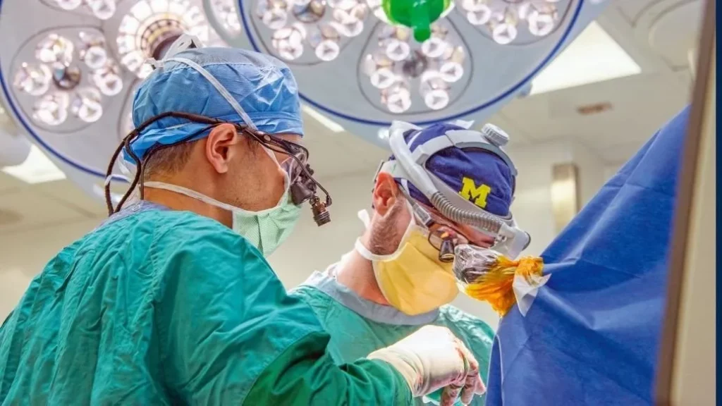
This technology represents the biggest advance in improving the detection rate of residual tumors in the last two decades. It could help drastically improve patients’ quality of life after surgery as it gains ground, and reduce the need for costly corrective surgery afterwards. It is also a prime example of Artificial Intelligence helping to improve patient outcomes in neurosurgery.
FastGlioma could also be expanded to help other types of patients in the near future. “In future studies, we will focus on applying the FastGlioma workflow to other cancers, including lung, prostate, breast, and head and neck cancers,” said Aditya S. Pandey, who is a co-author of the work for technology.
0 Comments